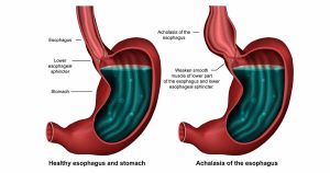
Overview
Diagnosis of Achalasia
Achalasia can be challenging to diagnose because its symptoms resemble other digestive disorders. A thorough evaluation typically includes:
-
Esophageal Manometry:
This test measures esophageal muscle contractions during swallowing and evaluates how effectively the lower esophageal sphincter (LES) relaxes. It is the most accurate test for confirming achalasia and determining its subtype. -
Barium Swallow X-rays:
After drinking a barium solution or swallowing a barium pill, X-rays visualize the esophagus, stomach, and upper intestine. The barium outlines the digestive tract, helping identify narrowing, blockages, or structural abnormalities. -
Upper Endoscopy (Esophagogastroduodenoscopy):
A flexible tube with a camera examines the esophagus, stomach, and duodenum. Endoscopy can detect partial obstructions, rule out malignancy, and obtain tissue biopsies to check for complications like Barrett’s esophagus. -
Functional Luminal Imaging Probe (FLIP) Technology:
FLIP is an advanced technique that measures esophageal distensibility and LES function, providing additional diagnostic information when traditional tests are inconclusive.
Treatment of Achalasia
The primary goal of treatment is to relax or widen the lower esophageal sphincter so that food and liquids can pass more easily into the stomach. Treatment choice depends on age, overall health, and disease severity.
Nonsurgical Treatment
-
Pneumatic Dilation:
A balloon is inserted endoscopically into the LES and inflated to enlarge the opening. Some patients require repeat dilation if the LES does not remain sufficiently open. About one-third of patients may need additional treatment within five years. Sedation is used during the procedure. -
Botulinum Toxin (Botox) Injection:
Botox can be injected into the LES during endoscopy to relax the muscle temporarily. Effects typically last up to six months, and repeated injections may complicate future surgery. Botox is mainly recommended for patients who are not candidates for dilation or surgery. -
Medications:
Muscle relaxants such as nitroglycerin or nifedipine may be prescribed before meals. These drugs provide limited benefit and may have significant side effects, so they are reserved for patients unsuitable for other interventions.
Surgical Treatment
-
Heller Myotomy:
This surgical procedure involves cutting the LES muscle to allow food to pass into the stomach. It can be performed laparoscopically, minimizing invasiveness. Postoperative gastroesophageal reflux disease (GERD) is a possible complication. -
Fundoplication:
Often performed with Heller myotomy to prevent reflux, fundoplication involves wrapping the top of the stomach around the lower esophagus to create an anti-reflux barrier. This procedure is usually done laparoscopically. -
Peroral Endoscopic Myotomy (POEM):
POEM is a minimally invasive endoscopic approach in which the LES muscle is cut from inside the esophagus. POEM may be combined with or followed by fundoplication or managed with acid-suppressing medications if reflux occurs.
Follow-up and Long-Term Management
-
Regular monitoring ensures proper swallowing function and detects complications such as GERD.
-
Lifestyle adjustments, including eating smaller meals and avoiding lying down immediately after eating, can support treatment outcomes.
-
Periodic imaging or endoscopic evaluation may be recommended in patients at risk for complications or symptom recurrence.
Advertisement