Overview
Diagnosis of Blocked Tear Duct
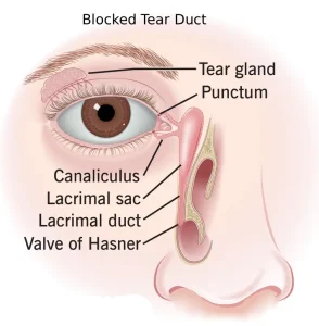
Diagnosing a blocked tear duct involves evaluating your symptoms, examining your eyes, and performing specialized tests.
-
Symptom Review and Eye Examination:
-
Your healthcare professional will ask about tearing, discharge, or recurrent eye infections.
-
The inside of your nose may be checked for structural changes contributing to blockage.
-
-
Tear Drainage Test:
-
A drop of special dye is placed on each eye to measure tear drainage.
-
If dye remains after five minutes, a blockage may be present.
-
-
Irrigation and Probing:
-
Saline may be flushed through the tear system, or a slender probe may be inserted through the puncta (tiny eyelid drainage openings) to locate or treat blockages.
-
-
Eye Imaging Tests:
-
Contrast dye is passed through the tear duct system.
-
X-ray, CT scan, or MRI can help pinpoint the location and cause of the blockage.
-
Treatment of Blocked Tear Duct
Treatment depends on the cause and severity of the blockage, and multiple approaches may be combined.
-
Medicines to Treat Infection:
-
Antibiotic eye drops or oral medications are prescribed if an infection is present.
-
-
Watch-and-Wait or Massage:
-
Many infants’ tear ducts improve naturally within the first months of life.
-
Gentle massage techniques may help open a thin tissue membrane covering the nasolacrimal duct.
-
Adults with facial injuries may wait for swelling to subside before treatment.
-
-
Dilation, Probing, and Flushing:
-
Infants: Done under general anesthesia; punctal openings are dilated and a thin probe is used.
-
Adults: Partial punctal narrowing may be treated with simple irrigation, often providing temporary relief.
-
-
Stenting (Intubation):
-
A thin tube is threaded through the puncta into the nose to keep the tear duct open.
-
Tubes are typically left in place for about three months.
-
Possible complications include inflammation.
-
-
Balloon Catheter Dilation:
-
Used if other treatments fail or blockage recurs.
-
A catheter with a balloon is threaded through the tear duct and inflated to open the passageway.
-
-
Tumor-Related Blockages:
-
If a tumor is causing the blockage, treatment targets the tumor, possibly including surgery or other therapies.
-
Surgery for Blocked Tear Duct
The most common surgical procedure is dacryocystorhinostomy (DAK-ree-oh-sis-toe-rye-nohs-tuh-me), which opens the tear drainage passage.
-
External Dacryocystorhinostomy:
-
A cut is made near the lacrimal sac to connect it to the nasal cavity.
-
A stent keeps the passageway open, and the incision is closed with stitches.
-
-
Endoscopic or Endonasal Dacryocystorhinostomy:
-
Instruments and a camera are inserted through the nasal opening.
-
No external incision or scar, but success rates may be slightly lower than the external approach.
-
-
Post-Surgery Care:
-
Nasal decongestant sprays and eye drops prevent infection and reduce inflammation.
-
Stents are usually removed after 6 to 12 weeks of healing.
-
Advertisement
