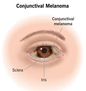
Overview
Diagnosis
Eye melanoma diagnosis usually begins with a comprehensive eye exam and may include several imaging and diagnostic tests to determine the size, location, and spread of the tumor.
-
Eye Exam:
A healthcare professional first checks the outside of the eye for enlarged blood vessels, which can indicate abnormal growth inside. Specialized tools are then used to view the interior of the eye.-
Binocular indirect ophthalmoscopy: Uses a bright light and lenses mounted on the examiner’s head to view the back of the eye.
-
Slit-lamp biomicroscopy: Uses a microscope and focused light beam to magnify and illuminate internal eye structures.
-
-
Fundus Photography:
Captures color images of the inner eye surface (fundus) to help detect or monitor melanoma over time. Techniques like fundus autofluorescence may highlight abnormal cells. -
Eye Ultrasound:
Uses high-frequency sound waves from a handheld device (transducer) placed on the eyelid or eye surface to create detailed images of the tumor. -
Eye Angiography:
A colored dye is injected into a vein in the arm, allowing special cameras to capture images of blood flow in eye vessels.-
Fluorescein angiography and indocyanine green angiography are common forms.
-
-
Optical Coherence Tomography (OCT):
Uses light waves to produce detailed cross-sectional images of the retina and uvea, showing the tumor’s position and structure. -
Biopsy:
While often not required for diagnosis, a biopsy may be performed during treatment to analyze cancer cell characteristics and confirm the type of melanoma. -
Tests for Cancer Spread:
To check if the melanoma has metastasized, tests may include:-
Blood tests
-
Liver function tests
-
Chest X-ray
-
Ultrasound
-
CT scan
-
MRI scan
-
PET scan
-
Treatment
Treatment for eye melanoma depends on the tumor’s size, location, spread, and overall health condition of the patient. Options include radiation, laser therapy, surgery, and advanced targeted or immunotherapy treatments.
-
Observation (Watchful Waiting):
Small, non-growing melanomas may simply be monitored regularly. If they enlarge or cause symptoms, treatment can begin later. -
Radiation Therapy:
-
Often used for small to medium-sized melanomas.
-
Brachytherapy (Plaque Radiotherapy): A small, radioactive plaque is stitched over the tumor for a few days to destroy cancer cells.
-
External Beam Radiation: Uses precise X-ray or proton beams from outside the body, given over multiple sessions.
-
-
Laser Therapy:
Uses focused laser light to damage tumor cells.-
Thermotherapy (Infrared Laser): Sometimes combined with radiation for improved results.
-
-
Photodynamic Therapy (PDT):
Combines a light-sensitive drug (photosensitizer) with a laser light to destroy smaller tumors while sparing healthy tissue. -
Surgery:
-
Local Tumor Removal: Removes the melanoma along with a small margin of healthy tissue.
-
Enucleation: In cases of large or invasive tumors, the entire eye is removed.
-
A prosthetic implant is later fitted for cosmetic and movement restoration.
-
-
-
Targeted Therapy:
Uses medicines that block specific molecules involved in cancer cell growth. Often used when melanoma spreads or surgery isn’t possible. -
Immunotherapy:
Boosts the body’s immune system to recognize and destroy cancer cells. It may be recommended when eye melanoma spreads or cannot be surgically removed.
Advertisement

