Overview
Diagnosis of Cerebral Cavernous Malformations (CCMs)
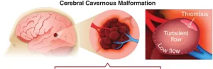
Cerebral cavernous malformations (CCMs) are often discovered accidentally during brain imaging for another condition. Many people with CCMs may not experience any symptoms, while others may develop neurological signs that lead to further testing.
Your neurologist will review your medical history, discuss any symptoms, and may recommend several diagnostic tests to confirm the presence of CCMs or rule out other conditions.
Common Diagnostic Tests for CCMs
1. Magnetic Resonance Imaging (MRI):
-
The most effective tool for diagnosing CCMs.
-
MRI provides detailed images of the brain or spinal cord, allowing doctors to locate and measure the malformation.
-
A contrast dye may be injected into a vein to enhance visibility.
2. Genetic Testing:
-
Recommended if you have a family history of CCMs.
-
Blood or saliva tests help identify gene mutations associated with inherited forms of the condition.
-
Genetic counseling can assist in understanding your risk and guide family members.
Specialized Care and Evaluation
At advanced neurological centers such as Mayo Clinic, a multidisciplinary team — including neurologists, neurosurgeons, and neuroradiologists — collaborates to design a personalized care plan.
They may use advanced imaging techniques like:
-
Functional MRI (fMRI): Measures brain activity and blood flow.
-
Tractography: Maps brain pathways to plan safer surgical procedures.
Treatment for Cerebral Cavernous Malformations (CCMs)
Treatment depends on the location, size, and symptoms of the malformation. Your healthcare team may suggest:
1. Observation (Watchful Waiting):
-
If your CCM isn’t causing symptoms, your doctor may recommend regular MRI scans to monitor for changes or bleeding.
2. Medications:
-
If seizures occur due to a CCM, anti-seizure medications may be prescribed to control them.
3. Surgery:
-
Recommended if the malformation causes repeated bleeding, seizures, or neurological issues.
-
The surgery involves removing the CCM to prevent further complications.
-
Advanced imaging (fMRI and tractography) helps make surgery more precise and safer.
Prognosis and Ongoing Research
The outlook for CCMs varies based on the number, size, and location of the lesions, and whether they cause symptoms or bleeding. Some CCMs remain stable for years, while others may progress.
Researchers are currently exploring non-surgical treatments — including medications that could lower the risk of bleeding — and new MRI techniques such as:
-
Quantitative Susceptibility Mapping (QSM)
-
Dynamic Contrast-Enhanced MRI (DCE-MRI)
These innovations aim to better predict disease progression and personalize treatment for each patient.
Key Takeaway
Early diagnosis through MRI and genetic testing is essential for managing cerebral cavernous malformations effectively. Treatment plans may range from observation to surgical removal, depending on symptoms and risk factors. Ongoing medical research continues to improve outcomes for those living with CCMs.
Advertisement
