Overview
Diagnosis
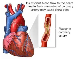
Chest pain does not always mean a heart attack, but because it can be life-threatening, emergency medical professionals first test for heart-related and serious lung conditions, such as a collapsed lung or a blood clot in the lung.
Immediate Tests
To quickly determine the cause of chest pain, the following tests are often done first:
-
Electrocardiogram (ECG or EKG): A quick test that shows how the heart is beating. It helps determine if a person has had or is having a heart attack. Sticky sensors are placed on the chest, arms, and legs, connected to a computer that records the heart’s electrical activity.
-
Blood tests: These check for specific proteins that leak into the blood after heart damage from a heart attack.
-
Chest X-ray: Provides images of the lungs, heart size, and shape. It can reveal pneumonia or a collapsed lung.
-
Computerized tomography (CT) scan: Creates cross-sectional images of the chest. It can detect a blood clot in the lung or an aortic dissection (tear in the artery wall).
Follow-up Tests
Depending on the results of initial tests, further evaluation may include:
-
Echocardiogram: Uses sound waves to produce moving images of the heart, showing how blood flows through heart chambers and valves.
-
CT coronary angiogram: Produces detailed images of the coronary arteries to check for blockages or narrowing.
-
Exercise stress test: Measures how the heart responds to exercise, either by walking on a treadmill or with medicines that simulate exercise effects.
-
Coronary catheterization: Detects blockages in the heart’s arteries. A thin tube is inserted through a blood vessel in the groin or wrist and guided to the heart. A dye is injected to highlight arteries on X-ray images.
Treatment
Treatment for chest pain depends on the underlying cause.
Medications
Common medicines used to manage chest pain include:
-
Nitroglycerin: Relaxes heart arteries to improve blood flow; often given under the tongue for suspected heart-related pain.
-
Blood pressure medicines: Help relax and widen blood vessels, easing heart-related pain.
-
Aspirin: Used when chest pain is suspected to be heart-related; it helps prevent blood clots but doesn’t relieve pain.
-
Clot-busting drugs (thrombolytics): Dissolve clots blocking blood flow during a heart attack.
-
Blood thinners: Prevent future clots in heart or lung arteries.
-
Acid-reducing medicines: Lower stomach acid in cases of heartburn-related chest pain.
-
Anti-anxiety medicines: Help manage chest pain related to panic attacks; therapy like cognitive behavioral therapy may also be recommended.
Surgical and Other Procedures
For serious causes of chest pain, other treatments may include:
-
Angioplasty and stent placement: A balloon-tipped tube opens a blocked heart artery, and a stent keeps it open to improve blood flow.
-
Coronary artery bypass graft surgery (CABG): Open-heart surgery where a vein or artery from another part of the body is used to bypass a blocked heart artery.
-
Emergency repair surgery: Urgent surgery to repair a ruptured aorta (aortic dissection), a life-threatening condition.
-
Lung reinflation: If the lung has collapsed, a chest tube may be inserted to help it expand again.
Request an appointment
Advertisement
