Overview
Diagnosis
Diagnosing a craniopharyngioma usually begins with a review of medical history and symptoms. Several tests may be used to confirm the diagnosis and understand the tumor’s effect on the brain.
-
Neurological exam: This includes testing vision, hearing, balance, coordination, reflexes, and growth and development. These results can show which area of the brain may be affected by the tumor.
-
Blood tests: Changes in hormone levels can suggest that the tumor is affecting the pituitary gland.
-
Imaging tests: X-rays, CT scans, and MRI scans provide detailed images of the brain to identify the size, location, and features of the tumor. In certain situations, additional imaging may be required.
Treatment
Treatment for craniopharyngioma often begins with surgery. The goal is to remove as much of the tumor as possible while protecting nearby brain structures. In some cases, radiation therapy, chemotherapy, or targeted therapy may also be used.
Surgery
Surgery is the primary treatment for most people with craniopharyngioma. The type of surgery depends on the tumor’s size and location.
-
Open craniopharyngioma surgery (craniotomy): The surgeon opens part of the skull to reach and remove the tumor.
-
Minimally invasive surgery (transsphenoidal procedure): Specialized tools are inserted through the nose to reach the tumor through natural passages, reducing the impact on the brain.
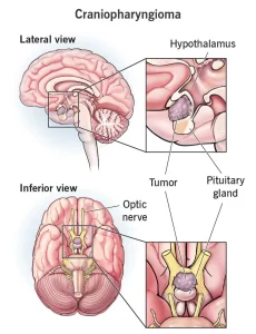
In some cases, it isn’t possible to remove the entire tumor safely due to its proximity to delicate structures such as the optic nerves and hypothalamus. Surgeons remove as much of the tumor as possible to preserve vision and brain function.
If the tumor causes hydrocephalus (fluid buildup in the brain), a tube may be placed to drain the fluid. This can be temporary or permanent. A permanent tube, called a shunt, drains the fluid to the abdomen.
Radiation therapy
Radiation therapy uses high-energy beams to destroy remaining tumor cells after surgery.
Types of radiation therapy include:
-
External beam radiation therapy: A machine directs precise beams of radiation toward the tumor while minimizing exposure to healthy tissue. Techniques such as proton beam therapy and intensity-modulated radiation therapy (IMRT) improve accuracy.
-
Stereotactic radiosurgery: A focused form of radiation that delivers multiple beams from different angles to the tumor, usually completed in one or a few sessions.
-
Brachytherapy: Radioactive material is placed inside or near the tumor to deliver radiation from within.
Chemotherapy
Chemotherapy may be used to kill tumor cells. For craniopharyngioma, it can sometimes be injected directly into the tumor, which helps target cancerous cells while limiting damage to nearby healthy tissue.
Treatment for papillary craniopharyngioma
Papillary craniopharyngioma is a rare form of the disease that often occurs in adults. Nearly all cases involve a DNA change known as the BRAF gene mutation.
Targeted therapy can be effective for this type of tumor. These medicines block specific signals that tumor cells use to grow and survive. Laboratory testing can determine if the tumor cells contain the BRAF mutation, guiding treatment decisions.
Advertisement
