Overview
Diagnosis
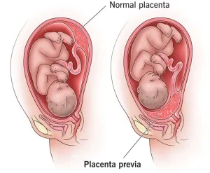
Placenta previa is diagnosed using ultrasound, either during a routine prenatal checkup or after an episode of vaginal bleeding. Most cases are identified during a second-trimester ultrasound.
The initial exam may use an abdominal ultrasound device. For more precise imaging, a transvaginal ultrasound may be performed, using a wandlike device inserted into the vagina. Care is taken to avoid disrupting the placenta or causing bleeding.
Treatment
If placenta previa is detected during a routine exam, more frequent ultrasound exams are usually scheduled to monitor changes in placental position.
In many cases, placenta previa resolves on its own as the pregnancy progresses. The uterus grows, the distance between the cervix and placenta may increase, and the edges of the placenta near the cervix may shrink. If resolved, vaginal delivery may be possible. If not, a C-section is planned.
Treatment of bleeding:
• Vaginal bleeding after 20 weeks is treated as a medical emergency. Hospital admission may be required for monitoring both mother and baby, and a blood transfusion may be needed.
• If bleeding occurs at or after 36 weeks, a C-section is typically performed. Emergency C-section may be necessary for severe bleeding or risk to mother or baby.
• If bleeding stops for at least 48 hours, you may be sent home, but repeated or heavy bleeding may require continued hospitalization.
Treatment with no bleeding:
To reduce the risk of bleeding, you may be advised to avoid:
• Sexual activity that could cause orgasm
• Moderate or strenuous exercise
• Moderate or heavy lifting
• Standing for long periods
Emergency care should be sought if vaginal bleeding or contractions occur. Support at home for transportation to a hospital may be necessary.
Planned C-section delivery:
Even without further bleeding, a C-section is usually scheduled between 36 and 37 weeks. If delivery is planned before 37 weeks, corticosteroids may be given to help the baby’s lungs develop.
Advertisement
