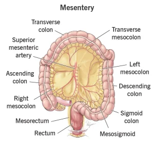
Overview
Diagnosis
Diagnosing sclerosing mesenteritis involves a combination of physical exams, imaging, and sometimes tissue sampling.
-
Physical exam — During a physical exam, your healthcare professional may feel a mass in the upper abdomen, which is a common sign of sclerosing mesenteritis.
-
Imaging tests — Abdominal imaging, such as CT scans or MRI, can help detect the presence of sclerosing mesenteritis.
-
Biopsy — A tissue sample may be taken to confirm the diagnosis and rule out other conditions, including certain cancers such as lymphoma or carcinoid. Biopsies can be collected during surgery or through a needle inserted into the skin.
A biopsy is particularly important before starting treatment to ensure an accurate diagnosis.
Treatment
Treatment for sclerosing mesenteritis depends on the severity of symptoms. If you are not experiencing discomfort, you may not need immediate treatment and may only require periodic imaging to monitor the condition.
Medicines
Medications are used to reduce inflammation and control scar tissue growth:
-
Corticosteroids — Medicines such as prednisone help control inflammation. They may be used alone but are often combined with other treatments. Corticosteroids are generally used for no longer than 3 to 4 months due to potential side effects.
-
Hormone therapy — Tamoxifen may slow scar tissue growth and is often combined with corticosteroids or other medicines. Tamoxifen can increase the risk of blood clots, so a daily aspirin may be prescribed. Progesterone may be used as an alternative but also carries significant side effects.
-
Other medicines — Additional treatments may include azathioprine, colchicine, cyclophosphamide, or thalidomide, depending on the case.
Surgery
Surgery may be required if scar tissue obstructs the digestive tract and prevents food from moving through properly.
Request an appointment
Advertisement
