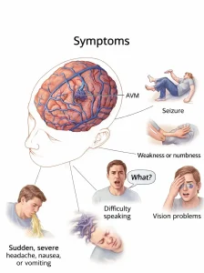
Overview
Diagnosis of Brain AVM
Your healthcare professional evaluates symptoms and conducts a physical exam. Imaging tests are usually performed by neuroradiologists, specialists in brain and nervous system imaging.
Tests used to diagnose brain AVMs include:
-
Cerebral angiography: The most detailed test for diagnosing a brain AVM. A catheter is inserted into an artery in the groin or wrist, guided to the brain using X-rays. Contrast dye highlights arteries and veins, helping plan treatment. Also called cerebral arteriography.
-
CT scan: Uses a series of X-rays to create cross-sectional images of the brain.
-
CT angiography: Dye may be injected to visualize arteries feeding the AVM and draining veins.
-
-
MRI: Uses magnets and radio waves to create detailed brain images, more sensitive than CT for subtle tissue changes.
-
MR angiography: Dye may be injected to assess blood circulation and AVM location.
-
Treatment of Brain AVM
The main goal of treatment is to prevent bleeding (hemorrhage) and control symptoms such as seizures or headaches. Treatment choice depends on age, health, and AVM size/location.
Medicines
-
Symptom management: Medicines may be prescribed for headaches or seizures caused by the AVM.
Surgical Options
-
Surgical removal (resection):
-
A portion of the skull is removed to access the AVM.
-
Using a high-powered microscope, the surgeon clips and removes the AVM.
-
The skull bone is reattached, and the incision is closed.
-
Typically recommended when the AVM can be removed safely with minimal risk.
-
-
Endovascular embolization:
-
A catheter is inserted into an artery in the leg or wrist and threaded to the brain AVM.
-
An embolizing agent (particles, glue, microcoils) is injected to block blood flow into the AVM.
-
Less invasive than traditional surgery; often used before other surgical procedures to reduce bleeding risk or AVM size.
-
-
Stereotactic radiosurgery (SRS):
-
Uses highly focused radiation beams to damage AVM vessels and cause scarring.
-
The AVM closes slowly over 1–3 years.
-
Ideal for small or hard-to-access AVMs, or those that haven’t caused hemorrhage.
-
Monitoring
-
Observation may be recommended for AVMs with few or no symptoms or located in difficult-to-treat brain areas.
-
Monitoring includes regular checkups and imaging with the healthcare team.
Potential Future Treatments
-
Research focus: Predicting hemorrhage risk using blood pressure within the AVM and hereditary syndromes.
-
Imaging innovations: 3D imaging, brain tract mapping, and functional imaging to improve surgical precision and safety.
-
Advances in surgery and embolization: New microsurgery, embolization, and radiosurgery techniques allow treatment of previously hard-to-access AVMs safely.
Advertisement

