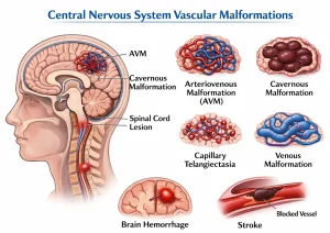
Overview
Diagnosis
To diagnose a central nervous system (CNS) vascular malformation, a healthcare professional reviews your medical and family history for stroke, epilepsy, or related conditions. A physical examination may also be performed. During this exam, your healthcare provider may listen to the blood flow in your arteries with a stethoscope. Some vascular malformations create a whooshing sound known as a bruit, caused by fast blood flow through the malformed vessels.
Imaging Tests
Imaging tests help detect and evaluate a central nervous system vascular malformation. A magnetic resonance angiogram (MRA) or a computerized tomography angiogram (CTA) may be used to identify the malformation and plan treatment.
An angiogram provides detailed images of blood flow through arteries or veins. A contrast material is injected into the bloodstream through an IV, allowing the blood vessels to be visible on the angiogram image.
Some malformations, such as cavernous malformations, can be detected using standard MRI or CT scans.
Treatment
Treatment for a central nervous system vascular malformation depends on factors such as:
-
The type of malformation
-
Its location
-
The symptoms it causes
-
The risk of bleeding
In some cases, regular monitoring may be recommended if the malformation poses little risk.
Medicines
Medications can help manage symptoms caused by venous malformations. These may include antiseizure medicines for seizures and pain relievers for headaches.
Surgery or Other Procedures
Certain central nervous system vascular malformations that carry a high risk of bleeding may require surgical removal. The appropriate procedure depends on the specific malformation.
Surgery involves opening the brain or spinal cord to remove the abnormal blood vessels. This approach is commonly used for small, accessible arteriovenous malformations. However, as with any surgery, risks include infection, bleeding, and potential damage to nearby healthy tissue.
Stereotactic radiosurgery is a noninvasive treatment that uses precisely targeted radiation beams to damage the vessel walls of the malformation, causing it to shrink over time. While this method has fewer risks than traditional surgery, there is still a chance of radiation affecting healthy tissue.
Endovascular embolization is another treatment option. A thin catheter is inserted into an artery in the leg or groin and guided to the malformation using X-ray imaging. Through this catheter, coils or a gluelike substance are released to block the blood flow to the malformation.
Embolization may be used alone or in combination with surgery. However, it may not completely remove the malformation, and its effects may not be permanent.
Advertisement
