Overview
Diagnosis
To diagnose Chiari malformation, your healthcare professional will review your medical history, discuss your symptoms, and perform a physical examination.
Imaging tests are essential to confirm the diagnosis and identify the cause. These may include:
-
Magnetic resonance imaging (MRI):
MRI is the primary test used to diagnose Chiari malformation. It uses powerful radio waves and magnets to produce detailed 3D images of the brain and spinal cord.-
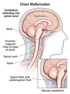
It helps detect structural differences in the brain, such as whether the cerebellum extends into the spinal canal.
-
MRI is a safe and painless test that can also be repeated over time to monitor the condition.
-
-
Computerized tomography (CT) scan:
A CT scan uses X-rays to create cross-sectional images of the body. It helps detect brain damage, tumors, bone or blood vessel problems, and other abnormalities.
More Information:
Brain magnetic resonance imaging
Brain CT scan
Treatment
Treatment for Chiari malformation depends on the severity of the condition and symptoms.
-
No symptoms:
If you don’t experience symptoms, your healthcare professional may recommend regular monitoring with exams and MRIs rather than active treatment. -
Pain management:
If headaches or pain are your main symptoms, pain-relief medicines may be prescribed to help manage discomfort.
Reducing Pressure with Surgery
When Chiari malformation causes symptoms, surgery is the most common and effective treatment.
The main goals of surgery are to:
-
Prevent further damage to the brain and spinal cord.
-
Relieve pressure and restore the normal flow of cerebrospinal fluid.
Common surgical procedures include:
-
Posterior fossa decompression:
This is the most frequent surgery for Chiari malformation. It involves removing a small portion of bone at the back of the skull to relieve pressure and create more space for the brain. -
Dura mater patching:
The dura mater (the brain’s protective covering) may be opened, and a patch — either from your own tissue or synthetic material — may be added to enlarge the space. -
Spinal decompression:
A small portion of the spinal column may be removed to reduce pressure on the spinal cord. -
Shunt placement:
If you have a syrinx (fluid-filled cavity in the spinal cord) or hydrocephalus (fluid buildup in the brain), a shunt may be inserted to drain excess fluid.
Surgical Risks and Follow-Up
As with any surgery, there are potential risks, including:
-
Infection
-
Fluid buildup in the brain
-
Cerebrospinal fluid leakage
-
Wound healing problems
Discuss the risks and benefits of surgery with your healthcare professional before proceeding.
Most people experience symptom relief after surgery. However, if nerve damage has already occurred, surgery may not reverse it.
Post-surgery care involves regular follow-up visits and imaging tests to monitor recovery and ensure normal cerebrospinal fluid flow.
Advertisement
