Overview
Diagnosis
Diagnosis of aortic coarctation depends on how severe the heart condition is. Severe cases are usually identified soon after birth and can sometimes be detected during pregnancy through ultrasound imaging.
Milder cases may not be found until later in childhood or adulthood.
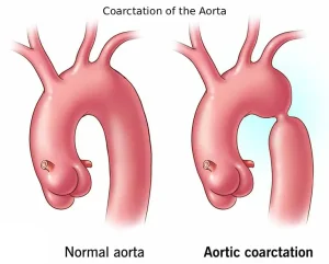
To diagnose aortic coarctation, a healthcare professional measures blood pressure in the arms and legs. When the aorta is narrowed, blood pressure tends to be higher in the arms and lower in the legs and ankles. The pulse in the legs may also feel weak or delayed. A heart murmur, which sounds like a whooshing noise, may be heard during examination.
Tests
Several tests help confirm the diagnosis and determine the severity of the narrowing:
-
Echocardiogram – Uses sound waves to create images of the beating heart, showing how blood flows through it. It identifies where and how much of the aorta is narrowed and helps plan treatment.
-
Electrocardiogram (ECG or EKG) – A quick, painless test that measures heart rhythm. Electrodes are attached to the chest, arms, or legs, and the results are displayed on a monitor or printed. Severe narrowing may cause thickening of the lower heart chambers.
-
Chest X-ray – Provides images of the heart and lungs and may show narrowing at the site of coarctation.
-
Cardiac MRI – Uses magnetic fields and radio waves to produce detailed images of the heart and blood vessels. It helps determine the location and extent of the narrowing and may guide treatment.
-
CT scan – Creates detailed cross-sectional images using X-rays to assess the structure of the heart and aorta.
-
Coronary angiogram with cardiac catheterization – A thin tube called a catheter is guided through a blood vessel, usually in the groin or wrist, to the heart. Dye is injected to make blood flow visible on X-ray images. This test helps measure the severity of the narrowing.
-
CT angiogram – Uses a dye and special X-rays to show how blood moves through the heart’s arteries and veins. It identifies the location and severity of the coarctation and whether other vessels are affected.
More Information
-
Coarctation of the aorta care at Mayo Clinic
-
Cardiac catheterization
-
Chest X-rays
Treatment
Treatment for aortic coarctation depends on the patient’s age at diagnosis and the severity of the narrowing. Treatment options may include:
-
Medicines
-
A heart procedure
-
Surgery
If other congenital heart defects are present, they are often repaired during the same procedure.
Medication
Medications used in aortic coarctation include:
-
Blood pressure medicines – Prescribed to control high blood pressure before surgery. Even after successful repair, some people may continue to need these medicines.
-
Medicine to keep the ductus arteriosus open – In newborns, a temporary opening called the ductus arteriosus connects the aorta and pulmonary artery. It usually closes shortly after birth, but medication can keep it open to maintain blood flow until surgery is performed.
Surgery or Other Procedures
Surgery or catheter-based procedures may be used to repair the narrowed section of the aorta. Common approaches include:
-
Balloon angioplasty and stenting – A catheter with a tiny balloon is used to widen the narrowed area. A small metal tube called a stent is often placed to keep the artery open and prevent renarrowing.
-
Resection with end-to-end anastomosis – The narrowed section of the aorta is surgically removed, and the two healthy ends are connected together.
-
Subclavian flap aortoplasty – A portion of the left subclavian artery, which supplies blood to the left arm, is used to enlarge the narrowed segment of the aorta.
-
Bypass graft repair – A synthetic tube called a graft is used to create a new pathway for blood flow around the narrowed part.
-
Patch aortoplasty – The narrowed section is cut open and expanded with a patch of material to widen the vessel, often used when a long segment of the aorta is involved.
After surgical repair, lifelong follow-up is essential to monitor blood pressure, detect complications, and ensure the aorta remains open and healthy.
Advertisement
