Overview
Diagnosis
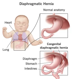
Congenital diaphragmatic hernia (CDH) is most often detected during a routine prenatal ultrasound. This imaging test uses sound waves to produce pictures of the uterus and developing baby. In some cases, CDH is diagnosed after birth or later in childhood if symptoms are mild or absent.
Healthcare professionals use ultrasound and other tests during pregnancy to monitor the growth and function of the baby’s lungs, heart, and other organs.
Prenatal ultrasound
The first fetal ultrasound usually takes place in the first trimester to confirm pregnancy and check for the number and size of the babies.
A second ultrasound, often between the fourth and sixth month, evaluates the baby’s development and the placement of internal organs. If CDH is suspected, additional ultrasounds may be scheduled to assess the severity of the condition and monitor its progression.
Other tests
Additional diagnostic tests may include:
-
Fetal MRI: Provides detailed images of the baby’s organs and tissues using magnetic fields and radio waves.
-
Fetal echocardiogram: Produces moving images of the baby’s heart to detect any structural or functional problems.
-
Genetic testing: Identifies chromosomal or genetic changes linked to CDH. Genetic counseling can help families understand results and possible outcomes.
Treatment
Treatment for congenital diaphragmatic hernia depends on when the condition is diagnosed and how severe it is. The healthcare team will develop a plan tailored to both the mother and baby.
Care before delivery
Expectant mothers with a CDH diagnosis typically undergo frequent ultrasounds and other monitoring tests.
An experimental approach called fetoscopic endoluminal tracheal occlusion (FETO) may be considered for severe cases. This prenatal surgery aims to help the baby’s lungs grow before birth.
FETO involves two steps:
-
Balloon placement: In the third trimester, a surgeon places a small balloon in the baby’s windpipe through a fetal endoscope. The balloon blocks fluid from escaping, encouraging lung growth.
-
Balloon removal: After 4 to 6 weeks, the balloon is removed so the baby can breathe after delivery.
If labor begins before the balloon can be removed, an ex utero intrapartum treatment (EXIT) procedure may be done. During this C-section, the placenta continues to provide oxygen until the balloon is removed and the baby is stabilized with a breathing tube.
FETO may not be suitable for all cases, and outcomes can vary. The healthcare team carefully evaluates each situation before recommending the procedure.
Care during delivery
Delivery can usually occur either vaginally or by C-section, depending on the mother’s and baby’s condition. The decision is made collaboratively between the mother and her healthcare provider.
Care after delivery
After birth, babies with CDH are typically treated in a newborn intensive care unit (NICU). Most require breathing support with a tube connected to a ventilator to assist lung and heart function.
Some infants with severe breathing issues may need extracorporeal membrane oxygenation (ECMO), a life-support technique that temporarily performs the work of the lungs and heart.
Surgery to close the hole in the diaphragm is usually necessary. The timing depends on the baby’s overall stability and lung function. Ongoing monitoring through chest X-rays ensures the repair remains intact.
After hospital discharge, some babies need:
-
Supplemental oxygen delivered through nasal prongs or a mask.
-
Feeding assistance to promote healthy growth.
-
Medications to manage related conditions such as acid reflux or pulmonary hypertension.
Regular follow-up visits are essential to track lung development, growth, and overall health, ensuring any complications are addressed early.
Advertisement
