Overview
Diagnosis
An eye care professional can diagnose dry macular degeneration by reviewing your medical and family history and performing a comprehensive eye exam. Several specialized tests may be used to confirm the diagnosis and determine the stage of the condition.
Examination of the back of the eye
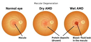
During this exam, eye drops are used to dilate the pupils. The eye doctor then examines the back of the eye with special instruments. A mottled appearance caused by yellow deposits under the retina, known as drusen, is a key sign of macular degeneration. People with the condition often have multiple drusen.
Amsler grid test
This simple test helps detect changes in the center of your visual field. You look at a grid of straight lines, and if you have macular degeneration, some of the lines may appear faded, broken, or distorted.
Fluorescein angiography
In this test, a fluorescent dye is injected into a vein in the arm. As the dye travels through the blood vessels in the eye, a special camera captures detailed images. These images can reveal retinal changes or abnormal blood vessel growth associated with macular degeneration.
Indocyanine green angiography
Similar to fluorescein angiography, this test uses another dye that helps highlight specific types of macular degeneration. It is sometimes performed together with fluorescein angiography for a more complete assessment.
Optical coherence tomography
This noninvasive imaging test provides detailed cross-sectional images of the retina. It helps identify areas of thinning, thickening, or swelling caused by fluid leakage from abnormal blood vessels beneath the retina.
Treatment
Although there is currently no way to reverse damage from dry macular degeneration, early detection and proactive care can help slow its progression. Lifestyle changes, vitamin supplements, and vision rehabilitation can help preserve remaining vision and improve quality of life.
Vitamin supplements
For people with intermediate or advanced macular degeneration, a specific combination of antioxidant vitamins and minerals has been shown to reduce the risk of further vision loss. Based on research from the Age-Related Eye Disease Study 2 (AREDS2), the recommended formulation includes:
-
500 mg of vitamin C
-
400 IU of vitamin E
-
10 mg of lutein
-
2 mg of zeaxanthin
-
80 mg of zinc as zinc oxide
-
2 mg of copper as cupric oxide
These supplements are not shown to benefit those with early-stage disease. Always consult your eye doctor before starting supplements to ensure they are suitable for you.
Low vision rehabilitation
Dry macular degeneration does not cause total blindness, but it affects central vision, which is essential for reading, driving, and recognizing faces. A low vision rehabilitation program can help you adjust and make the most of your remaining vision.
Rehabilitation may involve:
-
Working with a low vision specialist or occupational therapist
-
Using magnifying devices or special glasses
-
Learning new ways to perform daily tasks
-
Adapting your home and environment for better visibility
Surgery to implant a telescopic lens
For people with advanced dry macular degeneration in both eyes, surgery to implant a telescopic lens may be an option. The lens, which resembles a small plastic tube, magnifies the visual field and can enhance both near and distance vision.
While the telescopic lens offers improved central vision, it has a narrow field of view. It is most useful for specific situations, such as identifying street signs or objects at a distance in well-lit environments.
Advertisement
