Overview
Diagnosis
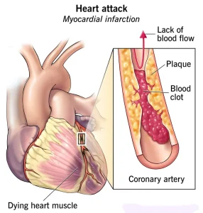
A heart attack is typically diagnosed in an emergency setting, where every minute matters. During regular checkups, healthcare providers may also screen for risk factors that can lead to a heart attack. When a heart attack occurs, immediate medical attention is critical to restore blood flow and minimize heart damage.
Diagnosis begins with checking vital signs such as blood pressure, pulse, and temperature, followed by a thorough medical history and symptom review. Various tests are used to confirm the diagnosis and assess the extent of heart damage.
Common diagnostic tests include:
-
Electrocardiogram (ECG or EKG): This is usually the first test done to detect a heart attack. It measures electrical signals in the heart to identify abnormalities in rhythm or heart damage.
-
Blood tests: Certain heart proteins, called cardiac markers, leak into the blood when the heart muscle is damaged. These markers help confirm a heart attack and measure its severity.
-
Chest X-ray: Provides an image of the heart and lungs to assess heart size and rule out other causes of chest pain.
-
Echocardiogram: Uses ultrasound waves to create moving images of the heart. It helps determine how well the heart is pumping and whether any areas are damaged.
-
Coronary catheterization (angiogram): A thin tube is guided through an artery to the heart, and dye is injected to visualize blockages in the coronary arteries.
-
Cardiac CT or MRI scans: These imaging tests produce detailed pictures of the heart to evaluate structure, blood flow, and the severity of damage.
Treatment
Immediate treatment is crucial during a heart attack to restore blood flow and prevent further heart damage. The specific treatment approach depends on whether the blockage in the artery is partial or complete. Oxygen therapy is usually given right away to improve oxygen supply to the heart.
Medications
Medications are often the first line of treatment during and after a heart attack:
-
Aspirin: Helps reduce blood clotting and maintain blood flow through narrowed arteries.
-
Clot busters (thrombolytics or fibrinolytics): These medications dissolve blood clots blocking the coronary arteries. The earlier they are given, the greater the benefit.
-
Other blood thinners: Drugs like heparin reduce blood stickiness and prevent new clots from forming.
-
Nitroglycerin: Relieves chest pain and improves blood flow by widening the blood vessels.
-
Morphine: Used to control severe chest pain that does not improve with nitroglycerin.
-
Beta blockers: Lower heart rate and blood pressure, reducing heart muscle damage and preventing further heart attacks.
-
ACE inhibitors: Help relax blood vessels, lower blood pressure, and reduce stress on the heart.
-
Statins: Lower bad cholesterol (LDL) to prevent future plaque buildup in the arteries.
Surgical and Other Procedures
Procedures may be performed to reopen blocked arteries and restore normal blood flow:
-
Coronary angioplasty and stenting (PCI): A catheter with a balloon at its tip is inserted into the blocked artery. The balloon is inflated to widen the artery, and a stent (a small mesh tube) is placed to keep it open. Some stents are drug-coated to prevent future narrowing.
-
Coronary artery bypass grafting (CABG): In this open-heart surgery, a healthy blood vessel from another part of the body is used to create a new pathway for blood to flow around the blocked artery. It may be done immediately after a heart attack or later, depending on the patient’s condition.
Cardiac Rehabilitation
After emergency treatment, cardiac rehabilitation plays a vital role in recovery. This structured program combines exercise, education, and lifestyle counseling to help improve heart health and prevent future heart problems.
Cardiac rehab typically includes:
-
Supervised exercise routines to strengthen the heart.
-
Guidance on a heart-healthy diet.
-
Stress management and emotional support.
-
Safe return to normal daily activities.
Participating in a cardiac rehab program significantly improves long-term outcomes, helping individuals recover faster, live longer, and lower the risk of another heart attack.
Advertisement
