Overview
Diagnosis
Inflammatory breast cancer is diagnosed through a clinical evaluation that includes a review of symptoms, medical history and a detailed breast exam. Additional tests help confirm the diagnosis and determine how far the cancer has spread.
Tests and procedures may include:
-
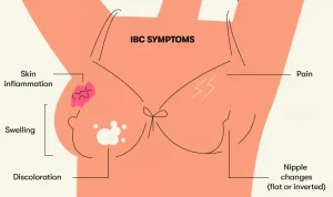
A physical exam to check for breast swelling, skin color changes and other signs of inflammatory breast cancer
-
Imaging tests such as a mammogram, ultrasound or breast MRI to look for abnormalities
-
A biopsy to remove a sample of breast tissue or skin cells for testing in a laboratory
Tests to determine the extent of cancer may be recommended after diagnosis. These tests help identify whether the cancer has spread to lymph nodes or other parts of the body. Imaging tests may include MRI, CT scans, bone scans or PET scans. Your healthcare professional will help decide which imaging tests are appropriate.
Inflammatory breast cancer is typically diagnosed at stages 3 or 4 because it grows quickly and spreads early. Stage 4 means the cancer has spread to other organs or bones.
Treatment
Treatment for inflammatory breast cancer usually begins with chemotherapy. Depending on how far the cancer has spread, surgery, radiation therapy and other treatments may also be recommended. These treatments aim to shrink the cancer, control its growth and improve long-term outcomes.
Chemotherapy
Chemotherapy uses powerful medicines to destroy cancer cells. These medicines may be given through a vein, taken as pills or both. For inflammatory breast cancer, chemotherapy is given before surgery. This is called neoadjuvant therapy and helps shrink the cancer to make surgery more effective.
If the cancer has a high chance of returning, additional chemotherapy may be recommended after other treatments are completed.
Surgery
After chemotherapy, surgery is usually performed to remove the affected breast and nearby lymph nodes. Surgery often includes:
-
Mastectomy to remove all breast tissue
-
Axillary lymph node dissection to remove lymph nodes under the arm
Breast reconstruction may be an option but is often delayed until all treatments are finished.
Radiation Therapy
Radiation therapy uses powerful energy beams to destroy remaining cancer cells after surgery. The radiation is directed at the chest, armpit and shoulder areas to reduce the risk of recurrence.
Targeted Therapy
Targeted therapy uses medicines that attack specific features within cancer cells. Some medicines focus on HER2, a protein that some breast cancer cells produce in excess. If your cancer is HER2 positive, targeted therapy may be given with chemotherapy and again after surgery.
Other targeted therapies may be available if the cancer spreads to other parts of the body. Testing your cancer cells helps determine which medicines may work best.
Hormone Therapy
Hormone therapy is used for cancers that rely on estrogen or progesterone to grow. This treatment may be recommended after surgery or if the cancer has spread. Options include:
-
Medicines that block hormones from attaching to cancer cells
-
Medicines that stop the body from producing estrogen
-
Treatments that stop hormone production by the ovaries
Immunotherapy
Immunotherapy helps the immune system recognize and destroy cancer cells. This treatment may be an option for triple-negative inflammatory breast cancer that has spread. Testing the cancer cells helps determine whether immunotherapy is likely to be effective.
Palliative Care
Palliative care focuses on improving quality of life during cancer treatment. It helps relieve pain, manage symptoms and provide emotional and practical support. This care can be given alongside treatments such as chemotherapy, surgery or radiation. People who receive palliative care along with standard cancer treatments often feel better and may live longer.
Advertisement
