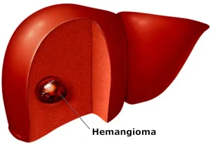
Overview
Diagnosis of Liver Hemangioma
Tests used to diagnose liver hemangiomas include:
Ultrasound
An imaging method that uses sound waves to produce images of the liver.
CT scan
A scan that combines a series of X-ray images taken from different angles and uses computer processing to create detailed images of the liver.
MRI scan
A technique that uses a magnetic field and radio waves to create detailed images of the liver.
Other tests may be used depending on your specific situation.
Treatment Options for Liver Hemangioma
If your liver hemangioma is small and causes no signs or symptoms, you likely will not need treatment. In most cases, liver hemangiomas never grow and never cause problems. Your doctor may schedule occasional follow-up exams to check whether the hemangioma is growing.
Treatment depends on the location and size of the hemangioma, whether you have more than one, your overall health, and your preferences.
Treatment options may include:
Surgery to remove the hemangioma
If the hemangioma can be easily separated from the liver, your doctor may recommend surgery to remove the mass.
Surgery to remove part of the liver
In some cases, a portion of the liver containing the hemangioma may need to be removed.
Procedures to stop blood flow to the hemangioma
Without blood supply, the hemangioma may stop growing or shrink.
- Hepatic artery ligation ties off the main artery feeding the hemangioma.
- Arterial embolization injects medicine into the artery to block it.
- Healthy liver tissue remains unaffected because it receives blood from other vessels.
Liver transplant surgery
In rare cases with a large hemangioma or multiple hemangiomas that cannot be treated by other methods, liver transplant surgery may be considered.
Radiation therapy
Radiation therapy uses powerful energy beams, such as X-rays, to damage the cells of the hemangioma. This treatment is rarely used because safer and more effective options are available.
Advertisement
