Overview
Diagnosis
To diagnose Paget’s disease of the breast, a healthcare professional often begins with a physical exam and a review of symptoms. Because most people with Paget’s disease of the breast also have cancer within the breast tissue, imaging tests are used to evaluate the breast for signs of cancer. Diagnosis is confirmed by removing a sample of tissue from the breast for testing in a lab.
Breast exam
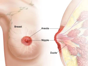
During a clinical breast exam, a healthcare professional looks at the breasts for any changes. For Paget’s disease of the breast, this can include changes in the skin or nipple. The healthcare professional then feels the breasts for lumps and checks along the collarbones and armpits for enlarged lymph nodes.
Mammogram
A mammogram is an X-ray of the breast used to screen for breast cancer. If a healthcare professional has concerns, a more detailed diagnostic mammogram may be ordered to closely examine both breasts.
During a mammogram, you stand in front of an X-ray machine designed for breast imaging. A technician places your breast on a platform and adjusts the equipment to match your height. Your head, arms and torso are positioned to get clear views of the breast tissue.
Breast ultrasound
Ultrasound uses sound waves to create images of structures inside the body. A breast ultrasound may help your healthcare team better understand a lump. It can show whether a lump is solid or fluid-filled, which helps determine the next steps in testing or treatment planning.
Breast magnetic resonance imaging
Magnetic resonance imaging, or MRI, uses a magnetic field and radio waves to create detailed images. A breast MRI may be used to check for additional areas of cancer in the affected breast or to look for cancer in the other breast. Before the scan, you typically receive an injection of dye to help the tissue show up more clearly on the images.
Biopsy
A biopsy removes a sample of tissue for laboratory testing. For Paget’s disease of the breast, this often includes taking a sample of skin from the affected nipple using a small cutting tool.
If imaging tests reveal a concerning area in the breast, a healthcare professional may remove a sample of breast tissue using a needle. Imaging such as X-ray or ultrasound helps guide the needle. Once the needle reaches the correct spot, tissue is removed for testing. A small marker may be placed at the biopsy site to help monitor the area in future imaging.
A core needle biopsy uses a long, hollow needle to remove a sample of tissue. The sample goes to a lab where pathologists examine it to determine whether cancer is present.
More Information
Breast biopsy
Breast MRI
Breast self-exam for breast awareness
Mammogram
MRI
Treatment
Treatment for Paget’s disease of the breast often includes surgery. Additional treatments may include radiation therapy, chemotherapy or hormone therapy. Treatment recommendations may depend on whether cancer is limited to the nipple or also present in the breast tissue.
Surgery
Operations used to treat Paget’s disease of the breast include:
-
Removing the breast cancer. Breast-conserving surgery, or lumpectomy, removes the cancer while keeping most of the breast. For Paget’s disease of the breast, the surgeon may remove the nipple, areola and any breast tissue that contains cancer. Most people who have this operation also receive radiation therapy.
-
Removing all breast tissue. A mastectomy removes all breast tissue, including the ducts, lobules, fatty tissue and some skin, including the nipple and areola. The most common type is total or simple mastectomy.
-
Removing a few lymph nodes. A sentinel node biopsy removes the first lymph nodes where cancer is likely to spread. If no cancer is found in those nodes, more lymph node removal typically is not needed.
-
Removing several lymph nodes. Axillary lymph node dissection removes multiple lymph nodes from the armpit. This may be recommended if imaging shows that cancer has spread to the lymph nodes or if cancer is found during a sentinel node biopsy.
-
Removing both breasts. Some people choose a contralateral prophylactic mastectomy to remove the healthy breast if they have a high risk of developing breast cancer. High risk may be due to family history or certain genetic changes.
Complications depend on the type of surgery but may include pain, bleeding and infection. Removing lymph nodes can increase the risk of arm swelling, called lymphedema.
Some people choose breast reconstruction after a mastectomy. Reconstruction restores breast shape using implants or tissue from another part of the body. This may be done at the same time as mastectomy or at a later date. Many people ask for a referral to a plastic surgeon before breast cancer surgery.
Other treatments
After surgery, additional treatments may be recommended to reduce the risk of the cancer returning. Treatment decisions depend on the type of cancer and whether it has spread.
Other treatments for Paget’s disease of the breast may include:
-
Radiation therapy
-
Chemotherapy
-
Hormone therapy
Advertisement
