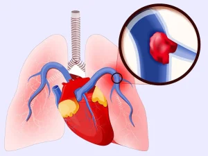
Overview
Diagnosis
A pulmonary embolism can be challenging to diagnose, especially when heart or lung conditions already exist. A health care provider typically reviews your medical history, performs a physical exam, and orders tests that may include the following options.
Blood tests
A blood test for the clot-dissolving substance D-dimer may be done. High levels can suggest the possibility of blood clots, though other factors may also raise D-dimer levels. Blood tests may also measure oxygen and carbon dioxide levels, since a clot in the lungs can reduce blood oxygen. Additional testing may check for inherited clotting disorders.
Chest X-ray
This noninvasive imaging test captures pictures of the heart and lungs. While an X-ray cannot diagnose a pulmonary embolism and may appear normal even when a clot is present, it helps rule out other conditions with similar symptoms.
Ultrasound
Duplex ultrasonography uses sound waves to examine veins for deep vein blood clots.
• It is commonly used for veins in the thigh, knee, calf, or sometimes the arms.
• A transducer moves across the skin, sending sound waves that create a moving image on a computer.
If no clots are seen, the likelihood of deep vein thrombosis is reduced. If clots are found, treatment usually begins right away.
CT pulmonary angiography
This CT scan generates detailed cross-sectional images to identify pulmonary embolisms within the lung arteries. Contrast dye may be injected through a vein during the scan to highlight the pulmonary arteries.
Ventilation-perfusion (V/Q) scan
A V/Q scan may be done when radiation or contrast exposure from a CT scan needs to be avoided. A small amount of radioactive tracer is injected to map blood flow and compare it with airflow in the lungs. This can help determine whether clots are contributing to symptoms of pulmonary hypertension.
Pulmonary angiogram
This is the most accurate test for diagnosing a pulmonary embolism, though it carries more risk and requires significant expertise.
• A catheter is inserted into a large vein, usually in the groin, and guided into the pulmonary arteries.
• Dye is injected while X-rays capture detailed images of blood flow.
Temporary heart rhythm changes or kidney risks from the dye may occur in some people.
MRI
MRI uses magnetic fields and radio waves to create detailed internal images. It is mainly used in pregnant individuals to avoid radiation exposure or in people whose kidneys could be affected by contrast dyes used in other imaging tests.
More Information
Chest X-rays
CT scan
MRI
Show more related information
Treatment
Treatment for pulmonary embolism aims to stop the clot from growing and prevent new clots. Immediate care is essential to avoid severe complications or death. Treatment may involve medication, procedures, and long-term management.
Medicines
Different medicines may be used, including blood thinners and clot-dissolving drugs.
Blood thinners
Anticoagulants help prevent existing clots from enlarging and stop new ones from forming while the body naturally breaks down clots. Heparin works quickly and may be given intravenously or by injection. It is often paired with an oral anticoagulant such as warfarin until the oral medicine becomes fully effective. Newer oral anticoagulants act faster and have fewer interactions, and some can be started without heparin. Bleeding is the most common side effect with all anticoagulants.
Clot dissolvers
Thrombolytics dissolve clots rapidly but can cause sudden, severe bleeding. Because of this risk, they are reserved for life-threatening situations.
Surgical and other procedures
Clot removal
A large, dangerous clot may be removed using a catheter inserted through the blood vessels.
Vein filter
A filter may be placed in the inferior vena cava to prevent clots from traveling to the lungs. This option is used when anticoagulants cannot be taken or if clots continue to form despite treatment. Some filters are removable once they are no longer needed.
Ongoing care
Because the risk of another deep vein thrombosis or pulmonary embolism remains, ongoing management is important.
• Continue anticoagulant therapy as directed.
• Attend regular follow-ups to monitor treatment and prevent complications.
Staying consistent with medical care helps reduce the likelihood of future clots and supports long-term health.
Advertisement
