Overview
Diagnosis
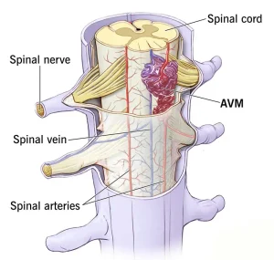
Spinal arteriovenous malformations can be challenging to diagnose because their symptoms often resemble those of other spinal disorders. These may include spinal dural arteriovenous fistula, spinal stenosis, multiple sclerosis, or spinal cord tumors. Because of this overlap, detailed evaluation and advanced imaging are usually required.
Your healthcare professional may recommend tests to rule out other possible causes of your symptoms.
Magnetic resonance imaging (MRI) uses strong magnets and radio waves to create detailed images of the spinal cord. A spinal MRI can show abnormal masses or changes caused by irregularly connected blood vessels associated with a spinal AVM.
Angiography is often necessary to clearly identify the location, structure, and blood flow of the abnormal vessels involved in a spinal AVM. During this procedure, a thin tube called a catheter is inserted into an artery in the groin and guided to the spinal cord. A special dye is injected into the spinal blood vessels, allowing them to be seen clearly using X-ray imaging.
Results from these imaging tests, along with neurological examinations and a review of symptoms, help confirm the diagnosis and guide treatment planning.
Treatment
Treatment for a spinal arteriovenous malformation may involve one or more approaches. Treatment aims to reduce symptoms, prevent worsening disability, and lower the risk of bleeding. The choice of treatment depends on factors such as the size, location, and blood flow of the AVM, along with your neurological exam findings and overall health.
The primary goal of treatment is to reduce the risk of hemorrhage and slow or stop the progression of neurological symptoms.
Medicines may be used to help manage symptoms such as back pain or stiffness. However, medicines alone do not treat the underlying AVM, and most spinal AVMs eventually require a procedural or surgical approach.
Surgery is often needed to remove or reduce the effects of a spinal AVM. There are several treatment options.
Conventional surgery involves making an incision to access and remove the AVM from surrounding tissue. Surgeons work carefully to avoid injury to the spinal cord and nearby structures. This approach is typically used when the AVM is relatively small and located in an area that is easier to reach.
Endovascular embolization is a minimally invasive procedure that can reduce blood flow to the AVM. A catheter is inserted into an artery in the leg and guided to the blood vessel supplying the AVM in the spinal cord. A gluelike substance or small particles are injected to block the artery and decrease blood flow. This method does not permanently destroy the AVM but can lower the risk of bleeding.
Endovascular embolization is sometimes performed before other types of surgery to reduce bleeding risk or make surgical removal more effective.
Radiosurgery uses precisely focused radiation to target the AVM. Over time, the abnormal blood vessels break down and close off. Radiosurgery is most commonly used for smaller AVMs that have not ruptured.
Because spinal AVMs are located close to the spinal cord, treatment is complex and requires careful planning. Your healthcare team will discuss potential benefits and risks of each option. Care from an experienced neurosurgeon and specialized medical team is essential for achieving the best possible outcome.
Advertisement
