Overview
Diagnosis
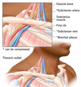
Diagnosing thoracic outlet syndrome (TOS) can be challenging because symptoms vary greatly among individuals. Healthcare professionals evaluate symptoms, medical history, and perform a physical exam. Imaging and other tests may be needed to confirm the diagnosis.
Physical exam: Your healthcare professional looks for signs such as a depression in the shoulder, a bony area above the collarbone, swelling, or color changes in the arm. Pulse and range of motion may be checked. Certain positions and movements may be used to reproduce symptoms, helping identify TOS.
Medical history: Discuss your symptoms, medical history, job duties, and physical activities with your healthcare professional.
Imaging and nerve study tests
Confirming TOS may require one or more of the following tests:
• Ultrasound. Uses sound waves to image the body and assess for venous or arterial TOS or other vascular conditions.
• X-ray. Detects an extra cervical rib and helps rule out other causes of symptoms.
• CT scan. Provides cross-sectional images; CT angiography with dye may show blood vessel compression.
• MRI. Uses magnetic fields and radio waves to create detailed images; sometimes dye is used. MRI can reveal anatomical differences like fibrous bands or cervical ribs that may compress vessels.
• Arteriography and venography. A catheter is inserted into arteries or veins to inject dye and capture X-ray images. This can detect compressed vessels or blood clots, and medications may be delivered through the catheter to dissolve clots.
• Electromyography (EMG). Needle electrodes assess muscle electrical activity at rest and during contraction to detect nerve damage.
Treatment
Early diagnosis often allows conservative treatment to be effective.
Physical therapy: First-line treatment for neurogenic TOS, focusing on strengthening and stretching shoulder muscles, improving posture and range of motion, and relieving pressure on nerves and blood vessels.
Medicines: Anti-inflammatory drugs, pain relievers, and muscle relaxants can reduce swelling, pain, and muscle tension. Blood-thinning medicines may be used if a clot is present.
Clot-dissolving medicines: Thrombolytics may be administered to dissolve blood clots in venous or arterial TOS. Anticoagulants may follow to prevent recurrence.
Injections: Local anesthetics, Botox, or steroid injections may help relieve pain in neurogenic TOS.
Surgical options: Surgery may be considered when conservative treatments are ineffective or symptoms persist. A thoracic or vascular surgeon performs thoracic outlet decompression, which may involve removing a muscle and part of the first rib to relieve compression. Surgery can also repair damaged blood vessels.
In venous or arterial TOS, clot removal or vessel repair may be done prior to decompression. Arterial replacement may use a graft from the patient or an artificial graft.
Surgery carries risks, including nerve injury, incomplete symptom relief, or recurrence of symptoms.
Advertisement
