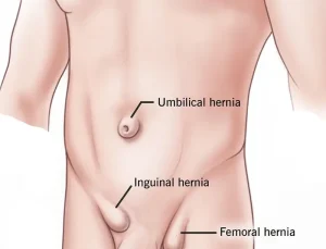
Overview
Diagnosis
An umbilical hernia is usually diagnosed during a physical examination. Your doctor can often identify the hernia by gently feeling the area around the bellybutton. In some cases, imaging tests may be recommended to check for complications or to confirm the diagnosis.
Common imaging tests that may be used include:
-
Abdominal ultrasound
-
X-ray
-
CT scan
These tests help evaluate the size of the hernia and detect any involvement of the intestines or other abdominal tissues.
Treatment
In most cases, umbilical hernias in babies close on their own by the age of 1 or 2. During a physical exam, a doctor may be able to gently push the bulge back into the abdomen. However, this should never be attempted at home, as it can cause harm.
It’s a myth that taping a coin or placing an object over the hernia can fix it. This method doesn’t work and may cause an infection by trapping germs under the tape.
Surgery for children is typically recommended if the hernia:
-
Is painful
-
Measures between 1/4 to 3/4 inch (1 to 2 centimeters) in diameter
-
Is large and doesn’t decrease in size over the first two years of life
-
Doesn’t close on its own by age 5
-
Becomes trapped or causes intestinal blockage
Surgery for adults is often advised to prevent complications, especially if the hernia becomes larger or painful.
During surgery, a small incision is made near the bellybutton. The herniated tissue is gently moved back into the abdominal cavity, and the opening in the abdominal wall is stitched closed. In adults, a surgical mesh may be used to reinforce the abdominal wall and reduce the risk of recurrence.
Advertisement

