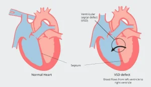
Overview
Diagnosis
Some ventricular septal defects are diagnosed soon after birth, while others may not be detected until later in life. In some cases, a VSD can be seen before birth during a pregnancy ultrasound.
If a ventricular septal defect is present, a healthcare provider may hear a whooshing sound, known as a heart murmur, when listening to the heart with a stethoscope.
Tests commonly used to diagnose a ventricular septal defect include:
-
Echocardiogram: The most common test used to diagnose VSD. Sound waves create moving images of the heart, showing how blood flows through the heart and its valves.
-
Electrocardiogram (ECG): This quick, painless test records the electrical activity of the heart to determine how fast or slow it’s beating.
-
Chest X-ray: This imaging test shows the condition of the heart and lungs. It can reveal an enlarged heart or extra fluid in the lungs.
-
Pulse oximetry: A fingertip sensor measures the amount of oxygen in the blood. Low oxygen levels may indicate a heart or lung problem.
-
Cardiac catheterization: A thin, flexible tube is inserted into a blood vessel and guided to the heart. This test helps doctors evaluate congenital heart defects and heart valve function.
-
Cardiac magnetic resonance imaging (MRI): Magnetic fields and radio waves create detailed heart images. This may be done if an echocardiogram doesn’t provide enough detail.
-
Computerized tomography (CT) scan: A CT scan uses X-rays to produce detailed images of the heart and may be used when further clarity is needed.
Treatment
Treatment for a ventricular septal defect depends on the size of the hole and the symptoms. Options may include regular monitoring, medications or surgery. Many small VSDs close on their own without the need for surgery.
For small VSDs, regular health checkups may be all that’s required. Medications may be used to manage symptoms, and some babies may need extra nutrition or oxygen if they tire easily during feeding.
Medications
While medications cannot close a ventricular septal defect, they can help manage symptoms and complications. Common treatments include:
-
Water pills (diuretics) to remove excess fluid and reduce strain on the heart
-
Oxygen therapy to improve oxygen levels in the blood
Surgeries or other procedures
Surgery may be needed if the defect is medium or large, or if it causes significant symptoms. Procedures include:
-
Open-heart surgery: The most common treatment for repairing VSDs. A surgeon uses stitches or a patch to close the hole between the heart’s lower chambers. This procedure requires a heart-lung machine and a chest incision.
-
Catheter procedure: A less invasive option for some VSDs. A catheter is guided through a blood vessel to the heart, and a small device is placed to close the defect without open-heart surgery.
After surgery or any repair procedure, lifelong follow-up care with a cardiologist is essential. Regular checkups and imaging tests help ensure that the heart continues to function well after treatment.
Advertisement

