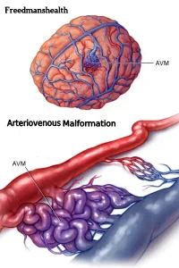
Overview
Diagnosis
To diagnose an arteriovenous malformation (AVM), a healthcare professional reviews your symptoms and conducts a physical examination.
Your provider may listen for a bruit, a whooshing sound caused by rapid blood flow through the arteries and veins of an AVM. This sound can sometimes affect hearing, disturb sleep, or cause emotional distress.
Tests commonly used to diagnose AVM include:
Cerebral angiography. Also called arteriography, this test uses a contrast dye injected into an artery to highlight blood vessels on X-rays, helping locate AVMs in the brain.
CT scan. Computed tomography scans help detect bleeding in the brain, head, or spinal cord using X-ray imaging.
CT angiography. Combines a CT scan with a contrast dye to detect bleeding or identify AVMs.
MRI. Magnetic resonance imaging uses strong magnets and radio waves to create detailed images of tissues, helping detect small structural changes in the brain or spinal cord.
Magnetic resonance angiography (MRA). This imaging method captures blood flow patterns, speed, and vessel structure to identify abnormal vessels in AVMs.
Transcranial Doppler ultrasound. Uses high-frequency sound waves to measure blood flow speed and detect AVMs, including identifying whether an AVM is bleeding.
Treatment
Treatment for an arteriovenous malformation depends on its location, size, symptoms, and risk of bleeding. Some AVMs may be carefully monitored with regular imaging if they have not caused bleeding or pose a low risk. Others may require active treatment.
When determining treatment, healthcare professionals consider:
-
Whether the AVM has bled.
-
Whether the AVM is causing symptoms like seizures, headaches, or neurological issues.
-
The location of the AVM and whether it can be safely treated.
-
The size and structure of the AVM.
Medicines. Medications can help control symptoms caused by AVMs, such as seizures, headaches, or pain.
Surgery. Surgical removal is often the main treatment for AVMs with a high risk of bleeding. Surgery is generally safer if the AVM is in an area where removal is unlikely to damage surrounding brain tissue.
Endovascular embolization. This minimally invasive procedure involves threading a catheter through arteries to the AVM. A substance is injected to block blood flow in the AVM. This procedure is often used before surgery or radiosurgery to reduce complications.
Stereotactic radiosurgery. This treatment uses highly focused radiation beams to target AVM vessels, damaging them and reducing blood flow over time.
Follow-up. After treatment or if an AVM is being monitored, regular imaging tests and checkups are necessary to ensure the AVM has not recurred and that there are no new complications.
Advertisement

